
Photography helps capture the world around us in hopes to share a moment with others – to stop and appreciate or investigate what is happening before us. These images help us stop and look within the frame of another’s view, so we can gain knowledge from a different perspective.
As time progresses, more and more upcoming technologies emerge that help us capture a clearer image. Oral health professionals embrace changing technologies when they capture the necessary images produced by dental x-rays. These x-rays help us gain information that would otherwise be unseen giving dentists the upper hand in treating your dental concerns.
Traditional x-ray photography
Your dentist uses x-ray photography extensively for taking images of your teeth, gums, and other invisible oral structures and conditions for providing you appropriate dental treatment. Traditional radiographic procedures used a chemically active plate as the sensor. The radiation passing through the examined tooth would impinge on the plate and affect the plate chemical in various locations to various extents depending on the radiation getting through. The plate was then chemically processed (in a wet process, normally called ‘washing’). The result was a dark plate which when held against light would show up an image. Areas which were more opaque to x-ray energy would show up as dark patches and those which passed x-rays more easily would show up brighter.
Digital X-ray photography
Modern technology has replaced the plate with electronic sensors which create the image immediately, doing away with the problems associated with the old (faithful) plate. The digital image is much like the image obtained in a modern digital camera- a soft image- that is, a set of ones and zeros, which is easily stored in any digital device.
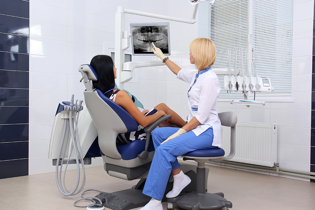
Dentist hand examining patient’s teeth X Ray
Types of Digital Dental Radiographs
- Digital dental radiographs are categorized as intraoral or extra-oral.
Intraoral x-rays- Intraoral x-ray photos are taken with the sensor placed inside the mouth against the tooth or teeth to be radiographed. The source is placed on the outside. The x-rays provide fine detail over a smaller area, e.g. a single tooth, and are the most commonly used type. - Bitewing x-rays- are taken with the patient biting on a thin plastic handle between his teeth. The handle holds the sensor vertically inside the mouth against the target area. This type of x-ray is useful when both upper and lower jaws must be viewed together, for example, in checking for occlusion problems.
- Periapical X-rays show a complete tooth from the crown to the root tips and the supporting bone. Unlike the bitewing a Periapical X-ray does not simultaneously show the upper and the lower teeth. It shows complete teeth (one or more) but of one jaw only.
- Extra-oral X-rays- the x-ray source and the sensor are both outside the mouth. They cannot provide the detail provided by intraoral X-rays and are not used for dealing with individual teeth, like root canal. Rather they are used to detect a wider scenario, e.g., monitor jaw development, temporomandibular problems, etc. Types of extra-oral X-rays include:
- Panoramic X-rays (Panorex) – the are also called OPG. The machine including the source and the sensor, rotates around the head, to capture the entire mouth, and all the teeth in each jaw, in one image. Panoramic X-rays are helpful in planning treatment for dental implants, jaw problems and impacted wisdom teeth, and also to diagnose bony cysts and tumors.
- Multi-slice computed tomography (MCT) reconstructs in detail a particular “slice” of the mouth while ignoring blurring all other layers. This type of X-ray is useful for examining structures that are difficult to see clearly.
- Cephalometric projections, can show the entire head, and help examine the teeth with reference to a patient’s jaw. A cephalometric projection is used by, orthodontists to develop their treatment plans.
- Sialography is used to examine problems with salivary glands, which may be a simple blockage, or Sjogren’s syndrome (an autoimmune disease that can reduce saliva and tear production). The glands are injected with a radiopaque dye as a contrast agent and then x-rayed.
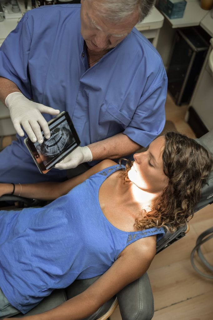
Dentist Office-Digital tablet with a patients x-rays
Cone beam computerized tomography (CBCT) or CT scans can show the interior structures as a three-dimensional image. The 3-D image is created by computer software from 2-D images taken from different aspects. CBCT is used to identify problems of facial bones like fractures or tumors. CT scans also are helpful in evaluating bone for dental implants and in difficult tooth extractions.
Benefits of Digital Dental Radiography
- The results are instant, and can be viewed on screen or printed. No chemical processing is required.
- Quick availability aids in patient comfort.
- Storage and retrieval is very convenient.
- Can be transmitted easily for review, comment, and lead to faster insurance settlements.
- During viewing the image can be magnified, rotated, contrast enhanced etc.
- As chemicals are avoided the digital imaging is eco-friendly.
- Digital x-rays normally demand less exposure to radiation.
In a world of technology several inventions and innovations allow for new groundbreaking discoveries. Whether you are capturing the perfect family memory or maintaining those pearly whites photography is key.